

A reader forwarded me a link to this new study
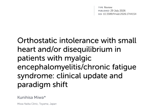
which included this nice diagram
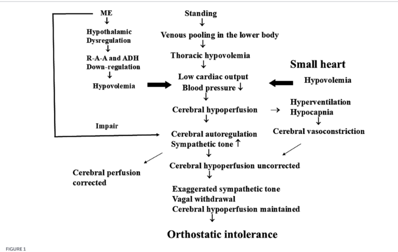
This is actually OLD news
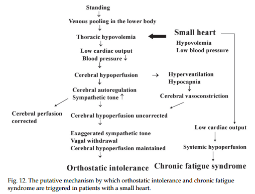
I covered this in a post from almost a decade ago, The Heart and Blood of the CFS Patient. This post cited studies from 2008. It also included this chart that bears great similarity to the above.
In new publications on ME/CFS, I too often seen results that were known and published in the past. Too often the prior studies were not cited — indicating that the researcher failed to do due diligence (or just search this site!!).
We have one factor that likely comes from DNA.
Inherited Coagulation Defects
This is another DNA variation that is over represented with ME/CFS and has been known for over a quarter century.
Addressing this was part of the plan that resulted in my remission.
Some more recent studies:
A notable 2024 plasma-proteomics study analyzed platelet-poor plasma from 15 people with ME/CFS and 10 non–SARS-CoV-2-infected controls. It found 24 proteins increased and 21 decreased in ME/CFS. Among the altered proteins were thrombospondin-1, platelet factor 4, and protein S—findings the authors interpreted as consistent with disrupted platelet/coagulation regulation and endothelial dysfunction. Complement proteins, including C9, were also lower. Its small sample size means this is hypothesis-generating rather than a confirmed biomarker panel.
A 2023 review in Blood Reviews concluded that multiple ME/CFS studies report platelet hyperactivation, anomalous clot formation, a more procoagulant phenotype, and endothelial dysfunction. It proposes impaired capillary-level exchange and tissue hypoperfusion as one possible connection to fatigue, cognitive symptoms, and exertional intolerance.
The “microclot” literature—especially studies using fluorescence methods, thromboelastography, and platelet imaging—has reported larger fibrinogen/amyloid-containing fibrinaloid deposits, platelet hyperactivation, and hypercoagulability in ME/CFS samples versus controls. These aggregates are hypothesized to be relatively resistant to normal fibrinolysis. However, assay standardization, replication by independent groups, and proof of clinical relevance remain important gaps
Myalgic Encephalomyelitis/Chronic Fatigue Syndrome: the biology of a neglected disease
Iron Levels — why females get ME/CFS more often?
This is more uncertain because ferritin has a complex interaction.
A 2023 Japanese study of people with Long COVID found those who went on to meet ME/CFS criteria had higher ferritin than both Long-COVID participants without ME/CFS and people without fatigue: median 193.0 µg/L versus 98.2 and 86.7 µg/L, respectively. The difference was particularly apparent in women, and ferritin correlated with fatigue severity measures.
Other Factors
The landmark DecodeME genome-wide association study compared 15,579 people with ME/CFS against 259,909 controls of European ancestry. Its initial 2025 results identified eight genome-wide significant genomic regions. The implicated biology clustered mainly around immune response to infection and nervous-system pathways.
Possible genetic clues to ME/chronic fatigue syndrome identified in massive study
Bottom Line
I view ME/CFS as a heterogeneous condition in which impaired oxygen delivery, oxygen utilization, or microcirculatory blood flow may contribute to a state of functional tissue hypoxia—meaning tissues may not receive or effectively use enough oxygen even when routine oxygen-saturation measurements appear normal.
In this model, a person may have pre-existing vulnerabilities that leave them relatively close to a physiological threshold. These vulnerabilities could include genetic predisposition, immune dysregulation, autonomic dysfunction, endothelial or microvascular abnormalities, mitochondrial or metabolic limitations, nutrient deficiencies, chronic inflammation, or prior infectious exposures. A triggering event—commonly an acute infection, but potentially another major physiological stressor—then pushes the system beyond its capacity to recover normally.
Once that threshold is crossed, the consequences may extend far beyond the initial trigger. Reduced perfusion or impaired cellular energy production could activate stress responses involving the autonomic nervous system, immune signaling, coagulation, oxidative stress, redox balance, metabolism, sleep regulation, and neuroinflammation. These systems interact with one another. For example, impaired tissue perfusion may worsen metabolic stress; metabolic stress may intensify inflammatory and autonomic signaling; and inflammation or vascular dysfunction may further impair blood flow and oxygen availability.
This could create a set of self-reinforcing feedback loops rather than one isolated defect. It may also help explain why people with ME/CFS can have normal results on many standard resting tests while developing profound symptoms after relatively minor physical, cognitive, or orthostatic exertion. Post-exertional malaise could reflect a delayed failure of recovery systems after demand exceeds an already limited physiological reserve.
Under this framework, correcting a contributor to hypoxia—such as iron deficiency, sleep-disordered breathing, low blood volume, impaired autonomic compensation, inflammation, or vascular dysfunction—may still meaningfully reduce symptoms. But it may be insufficient by itself to produce full recovery once downstream processes have become established. Recovery may require interrupting several interacting loops and allowing time for vascular, immune, metabolic, neurologic, and autonomic regulation to normalize.
This is a conceptual model rather than an established unified explanation for all ME/CFS. ME/CFS is likely biologically diverse: tissue hypoxia may be central in some people, secondary in others, and less important in still others.

