Dealing with neurologists …

I have had to deal with a neurologist — fortunately I walked into the appointment with a SPECT scan that a radiologist had read as early Alzheimer’s (I was having severe short term memory issues in the CFS flare when the SPECT was done). The session went well, he confirmed that the Alzheimer’s diagnosis was wrong and fortunately was familiar with CFS patients and deem it to be very real.

A reader wrote today

Hello, could you please link the research that shows which masses of the brain loose brain matter. I need to show this to my neurologist or she won’t help me get a spect done 😦 “

Pub Med Studies that should interest Neurologists

This is a technical post intended for neurologists, not brain fogged CFS patients.

central_nervous_system-smFrom Lactate in the brain by ME Reseach UK

  • “Mean lateral ventricular lactate concentrations measured by (1)H MRSI in CFS were increased by 297% compared with those in GAD (P < 0.001) and by 348% compared with those in healthy volunteers (P < 0.001), even after controlling for ventricular volume, which did not differ significantly between the groups. Regression analysis revealed that diagnosis accounted for 43% of the variance in ventricular lactate. CFS is associated with significantly raised concentrations of ventricular lactate, potentially consistent with recent evidence of decreased cortical blood flow, secondary mitochondrial dysfunction, and/or oxidative stress abnormalities in the disorder." [2009]
  • “Ventricular cerebrospinal fluid  lactate was significantly elevated in CFS compared to healthy volunteers, replicating the major result of our previous study. Ventricular lactate measures in MDD did not differ from those in either CFS or healthy volunteers. ” [2010]
  • ” We found elevated ventricular lactate and decreased GSH in patients with CFS and MDD relative to HVs. GSH did not differ significantly between the two patient groups. In addition, we found lower rCBF in the left anterior cingulate cortex and the right lingual gyrus in patients with CFS relative to HVs, but rCBF did not differ between those with CFS and MDD. We found no differences between the three groups in terms of any high-energy phosphate metabolites. In exploratory correlation analyses, we found that levels of ventricular lactate and cortical GSH were inversely correlated, and significantly associated with several key indices of physical health and disability. Collectively, the results of this third independent study support a pathophysiological model of CFS in which increased oxidative stress may play a key role in CFS etiopathophysiology.” [2012]
  • “Less efficient and costly processes of frontal cortex in childhood chronic fatigue syndrome. [2015]
    • “We conducted a study using a dual verbal task to assess allocation of attentional resources to two simultaneous activities (picking out vowels and reading for story comprehension) and functional magnetic resonance imaging. Patients exhibited a much larger area of activation, recruiting additional frontal areas. The right middle frontal gyrus (MFG), which is included in the dorsolateral prefrontal cortex, of CCFS patients was specifically activated in both the single and dual tasks; this activation level was positively correlated with motivation scores for the tasks and accuracy of story comprehension. In addition, in patients, the dorsal anterior cingulate gyrus (dACC) and left MFG were activated only in the dual task, and activation levels of the dACC and left MFG were positively associated with the motivation and fatigue scores, respectively. Patients with CCFS exhibited a wider area of activated frontal regions related to attentional resources in order to increase their poorer task performance with massive mental effort.”
    • scan

MRI:

    • “This study investigated responses to acute tryptophan feeding (after administration of 30 mg/kg body mass) using functional magnetic resonance imaging to investigate neural correlates of central fatigue during a cognitively demanding exercise, the counting Stroop task. Thus, tryptophan administration before the Stroop task caused distributed functional changes in primary sensory and in multimodal neocortex, including changes in a brain region, the activity of which has been shown previously to vary with conscious awareness (precuneus). Previous reports suggest that primary mechanisms of central fatigue may be predominantly subcortical. The present results demonstrate that neocortical activity changes are also found” [2007]
    • “The primary purpose of the study was to use functional magnetic resonance imaging (fMRI) to determine Results showed that mental fatigue was significantly related to brain activity during the fatiguing cognitive task but not the finger tapping or simple auditory monitoring tasks. Significant (p< or =0.005) positive relationships were found for cerebellar, temporal, cingulate and frontal regions. A significant (p=0.001) negative relationship was found for the left posterior parietal cortex. CFS participants did not differ from controls for either finger tapping or auditory monitoring tasks, but exhibited significantly greater activity in several cortical and subcortical regions during the fatiguing cognitive task." [2007]
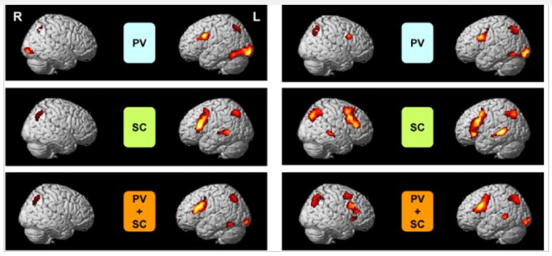
    • “In a series of two Blood Oxygen Level Dependent (BOLD) functional Magnetic Resonance Imaging (fMRI) studies, … Within and between regions of interest (ROI), group analyses were performed for both studies with statistical parametric mapping (SPM99). Findings showed that individuals with CFS are able to process challenging auditory information as accurately as Controls but utilize more extensive regions of the network associated with the verbal WM system. Individuals with CFS appear to have to exert greater effort to process auditory information as effectively as demographically similar healthy adults.” [2005]
    • Right arcuate fasciculus abnormality in chronic fatigue [2015]
      • “Bilateral white matter atrophy is present in CFS. …Right hemispheric increased FA may reflect degeneration of crossing fibers or strengthening of short-range fibers. Right anterior arcuate FA may serve as a biomarker for CFS.”


SPECT:

  • “Radiological imaging studies (SPECT, Xe-CT, and MRS) revealed decreased blood flow in the frontal and thalamic areas, and accumulation of choline in the frontal lobe.” [Learning and memorization impairment in childhood chronic fatigue syndrome manifesting as school phobia in Japan. 2004]
  • ” Regional blood flow studies by single photon-emission computerized tomography (SPECT) have been more consistent. They have revealed blood flow reductions in many regions, especially in the hind brain. Similar lesions have been reported after poliomyelitis and in multiple sclerosis–in both of which conditions chronic fatigue is characteristically present. In the well-known post-polio fatigue syndrome, lesions predominate in the RAS of the brain stem. ” [Chronic fatigue syndrome–aetiological aspects. 1997]
  • ” Patients with chronic fatigue syndrome had significantly more defects throughout the cerebral cortex on SPECT scans than did normal subjects (7.31 vs 0.43 defects per subject, p < .001). SPECT abnormalities were present in 13 (81%) of 16 patients, vs three (21%) of 14 control subjects (p < .01). SPECT scans showed significantly more abnormalities than did MR scans in patients with chronic fatigue syndrome(p < .025).” [Detection of intracranial abnormalities in patients with chronic fatigue syndrome: comparison of MR imaging and SPECT. 1994]
  • “Compared with the Normal Control  group, the CFS group showed significantly lower cortical/cerebellar rCBF ratios, throughout multiple brain regions (P < 0.05). Forty-eight CFS subjects (80%) showed at least one or more rCBF ratios significantly less than normal values. The major cerebral regions involved were frontal (38 cases, 63%), temporal (21 cases, 35%), parietal (32 cases, 53%) and occipital lobes (23 cases, 38%). The rCBF ratios of basal ganglia (24 cases, 40%) were also reduced.” [Assessment of regional cerebral perfusion by 99Tcm-HMPAO SPECT in chronic fatigue syndrome. 1992]

Brain Matter Volume

These was the requested studies asked for by the reader. I cite the 2015 studies that links to six earlier studies.

  • Gray matter volumes in patients with chronic fatigue syndrome [2015].
    • “Although most studies found Gray Matter volumes reduced in some brain regions in CFS, as we discussed above, there are so many factors that could affect brain structure should have been considered. So far, no study has controlled all these factors.”
    • We found 6 studies focusing on the change of GM volumes. Okada et al. [34] found in a voxel-based morphometric study of CFS that GM volumes reduced in bilateral prefrontal and that the affected areas extended from Brodmann area (BA) 8 to 9 in right cerebral hemisphere and from BA 9 to 11 in left. Puri et al. [35] also found that GM declined in occipital lobe, right angular gyrus, and posterior division of the left parahippocampal gyrus in a voxel-based morphometric 3-T MRI study. de Lange et al. [36] found that global GM volumes reduced by 8% compared to healthy controls and the rate of decline is 2.2 mL/year in a cohort study; moreover, the GM volumes could increase after cognitive behavioral therapy [37].

Bottom Line

CFS/ME alters the brain in many ways. Many of the alterations correspond to symptoms.

Melatonin – what do we know?

Melatonin has often been mentioned in my posts, without a deep dive into it.

Melatonin, also known as N-acetyl-5-methoxy tryptamine,[1] is a hormone that is produced by the pineal gland in animals and regulates sleep and wakefulness” [Wikipedia]

The more likely cause of sleep disorders with CFS/FM/IBS is hypoxia (low oxygen) which is known to cause severe sleep problems. Melatonin can help with that, but other oxygen delivery items should be considered too.

On the flip side, there some some bacteria that are associated with a lack of sleep: Partial Sleep Deprivation. On DataPunk.Net we read:

ENHANCES: 

INHIBITS: 

In other words, it contributes to a shift of the firmicutes/bacteroidetes ratio.

  • ” in particular through [Melatonin] ability to decrease the Firmicutes-to-Bacteroidetes ratio and increase the abundance of mucin-degrading bacteria Akkermansia, which is associated with healthy mucosa. ” [2017] – Checking Akkermansia levels first (most are low, but a few are high).


Bottom Line

If someone has a high firmicutes/bacteroidetes ratio (i.e.  firmicutes > 1.2x and /or bacteroidetes < .8x) then melatonin is a reasonable supplement, especially if there is overgrowth of  Coriobacteriaceae or Erysipelotrichaceae.  This applies to a subset of CFS patients.

Example:

f

On the Family Level

f2

The dosage is unclear. The literature reports: “0.1 mg to 50 mg/kg “, so for 100 lb individual:

  • 4.5 mg/day to
  • 2 gram/day

“For trouble falling asleep: 0.3 to 5 mg of melatonin daily for up to 9 months has been used. For sleeping problems in people with sleep-wake cycle disturbances: 2 mg to 12 mg taken at bedtime for up to 4 weeks has been used.” [WebMD]

Poison Control reports:

pc

“307 articles were elicited and 9 were related to Melatonin adverse effects. The range of MLT dose involved in the adverse reactions oscillated between 1 mg and 36 mg. The adverse reactions were: one patient with autoimmune hepatitis, one case of confusion due to MLT overdose, one case of optic neuropathy, four subjects with fragmented sleep, one psychotic episode, one case of nistagmus, four cases of seizures, one case of headache and two cases of skin eruptions.” [2001]

As always, consult with your medical professional before starting or altering your supplements.


Review of Drs. Naviaux and Davis and Suramin Work

A reader forwarded me a transcript of a conversation with Dr. Naviaux [Sep 13, 2017]. There are many points of interest raised so I thought that I would do a review.

For background on the Drs, see these MEpedia links

What is Suramin?

Suramin is a treatment for African sleeping sickness and river blindness. It is being tested for Zika virus.

What is Purinergic Sugnalling?

” form of extracellular signalling mediated by purine nucleotides and nucleosides such as adenosine and ATP.” [wikipedia]

What is adenosine is a mononucleoside consisting of adenine and D-ribose.[source]

Comments on Transcript

“A major misconception in medicine is that diseases that are “caused by different triggers are different diseases”. I believe this is wrong. The vast majority of chronic diseases are caused by the body’s response to stress or injury, and not the initial injury itself. In other words, over half of all chronic disease is caused by blocks in the healing process caused
by pathological persistence of the cell danger response (CDR). This is a categorically different way of thinking. It is different from the way doctors are trained to think about treating “acute” diseases like a strep infection, a broken leg, or a heart attack. “Chronic” disease needs to be analyzed and treated in a completely different way.”

  • absolutely agreed, for one class of these diseases it is the persistence of a microbiome dysfunction which presents as a metabolic disorder with metabolic abnormalities. Why — the microbiome is the producer of many metabolites.

“The most important medical decision is to determine:

  • if the person has an active infection or not. If yes, this must be treated.
  • If there is not an active infection, then we also need to determine if they have a household or occupational exposure to toxins that is ongoing.
  • If the answer to both investigations is “no”, then the patients would be eligible for the first pilot study.
  • Since our hypothesis is that antipurinergic therapy with suramin will treat the root problem of ME/CFS that is common to all patients, then you could say that the hypothesis we are testing in the first small pilot study of 10 patients, is that purinergic signaling abnormalities are common to ALL patients with ME/CFS, regardless of any “subset” they might belong to.”

Absolutely agreed for the first steps — the key is confirms actual infections with reliable tests (most Lyme tests do not meet that criteria); the second item should include oral/dental issues.

Bacteria Aspect

D-Ribose is produced by Bacillus subtilis which according to DataPunk, clusters with (seen with), Lactococcus lactis, Bacillus clausii, Lactobacillys plantarum and others. The bacillus genus also produces: Norepinephrine, Dopamine and Vitamin B12

Adenine (once known as Vitamin B4) is produced by E.Coli [1975]

In short: purinergic signaling abnormalities may be associated with microbiome changes.

The cost Aspects of doing CFS Studies..

“I estimate the CFS1 study of N = 10 patients will cost about $400,000…. so the cost of the clinical trials has nothing to do with the cost of the drug. Typically, it takes 15-30 people
on the clinical investigation team at each medical center to conduct a Phase III study. The main cost of the clinical trial is the salaries of the staff…the total cost of suramin drug development, from Phase I/II to Phase III trial is $20-$30 million.

What do we know about how Suramin impacts the microbiome?

Bottom Line

One of my frequent quotes is “They got the treatment right, but the theory behind may be incorrect”.  The drug, suramin, that they are wishing to test based on their hypothesis/theory does not exclude the hypothesis of a persistence microbiome dysfunction if they are successful. While information is sparse on the microbiome impact of suramin, the fact that it impacts candida indicates that it very capable of microbiome changes.


Beware nullifying probiotics by other “good stuff”

To illustrate the importance of being aware of interactions (which unfortunately can require retention levels above many CFS cognitive levels 😦 ) Consider the following:

A reader wrote this morning:

” The doctor has just prescribed mutaflor and ampicillin.”

We would love to believe that MDs, NDs, etc have complete understanding.

The Reality

I went to Mutaflor site and read:

“Antibiotic sensitivity of E. coli strain Nissle 1917

As a bacterial isolate from the pre-antibiotic era, E. coli strain Nissle 1917 (Mutaflor) is sensitive to all antibiotics  which are predominately directed against Gram-negative bacteria, e. g. coli bacteria.

A certain loss of ­Mutaflor-efficacy must be taken into account. Nevertheless, a protective ­Mutaflor-therapy should be initiated in addition to antibiotic therapy.

– Amikacin, [aminoglycoside]
— Amoxicillin/Clavulanic acid, [penicillin group of beta-lactam antibiotics]
— Ampicillin [penicillin group of beta-lactam antibiotics]
— Azlocillin [acylampicillin]
– Cefaclor [cephalosporin]
— Cefazolin [cephalosporin]
— Cefoperazone [cephalosporin]
— Cefotaxime [cephalosporin]
— Ceftriaxone [cephalosporin]
— Cephalotin [cephalosporin]
–Chloramphenicol [Amphenicol]
–Ciprofloxacin [quinolone]
– Doxycycline [tetracycline]
– Gentamicin [aminoglycoside]
– Latamoxef [penicillin beta-lactam]
– Mezlocilin [penicillin beta-lactam]
– Nitrofurantoin [macrolide]
— Norfloxacin [quinolone]
– Pipemidic acid [fluoroquinolones],
— Piperacillin [penicillin]
– Tetracycline [tetracycline],
— Ticarcillin [carboxypenicillin],
– Tobramycin [ aminoglycoside]
– Trimethoprim [Folic acid inhibitor]
– Sulfamethoxazole [sulfonamide]

Combination with antibiotics – natural antibiotic resistance of E. coli strain Nissle 1917

Mutaflor is not sensitive to antibiotics which are predominately directed against Gram-positive bacteria. That is why EcN may be combined with these ­antibiotics.

– Cefsulodin, Clindamycin [lincosamide]
– Erythromycin [macrolide]
– Metronidazole [Nitroimidazole]
– Penicillin G [Penicillin]
– Quinupristin/Dalfopristin [streptogramins]
– Rifampin [rifamycin]
– Teicoplanin [glycopeptide]
– Vancomycin [glycopeptide]”

Bottom Line

Trying to restore E.Coli has two probiotics available:  Mutaflor (covered above) and Symbioflor-2 . Both are known to take up residency and at least 1 is known to be human sourced. Studies found that Symbioflor-2 appears to have the same sensitivity [2008].

If antibiotics are taken, they should be done before E.Coli probiotics if one of the top list is prescribed. Antibiotics on the bottom list can be taken with E.Coli Probiotics. Personally, I suspect a cocktail of Metronidazole, Erythromycin and an E.Coli probiotic may play a similar role to CFS/IBS/SIBO etc as combination drug therapy does for H. Pylori infections.

Lactobacillus probiotics also kill off E.Coli!

The same issue exists with herbs and spices.  a rough way to get some guidance is to type the name of the herb/spice and “Gram-negative bacteria” (which E.Coli is)

search1

search2


Your mouth can trigger flares

The microbiome includes the mouth — and the bacteria in the mouth can repopulate the gut with bad bacteria. I have done several posts on the important of oral health. Today, a reader sent his experience using one of my suggestions: mastic gum. It is a interesting account:

“Hello Ken,

I’ve been taking your recommendation of chewing and swallowing mastic gum and to my amazement I seem to be getting my appetite back. It’s like the feeling of an empty stomach is back for me after 2 years. What else I discovered from chewing mastic gum was that is also seemed to have triggered my cfs symptoms again in an erratic way.


Now while this may seem paradoxical, I’ve looked into this further and found that the times when chewing gum made my cfs worse was specifically when I used the gum right against my back wisdom tooth which has significant decay under it. It seems like whenever I chew my gum anywhere else my energy and willpower stay constant, while if I chew the mastic gum over my wisdom tooth I have my energy diminish. I’ve trialed this multiple times, and am doing so right now as well. I can chew the mastic gum anywhere over my mouth and still feel fine, but when it gets into hard contact with my decaying wisdom tooth, my cfs symptoms re-emerge.


This is a critical piece of the puzzle for me, as I now know the amalgam likely created the environment for the infectious bacteria to flourish in my mouth, and now with the amalgam gone, the bacteria no longer stay thriving as they once used to, but they are still there lurking, and certain triggers may be able to bring them back into a larger presence within my microbiome.


As my decaying wisdom tooth is on the same side my amalgam once was. I believe I got 100% of the old amalgam out on my second dental visit, along with the tooth being cleared out. However the surrounding teeth are still likely to be suspect, and my experience with mastic gum proves it.”


For myself, mastic gum caused ‘night-time dry mouth’ to disappear.

Reference posts:

Bottom Line

“Your mileage may vary” – that is. different results from above may occur for different people.  I usually chew mastic gum with a little myrrh  and frankincense (boswellia sap) — the latter two are an ‘acquired taste’.