CFS/ME Research and the microbiome

A reader forwarded a new study from March, 2021. Unfortunately, ME/CFS seems to be in an infinite loop of similar hypotheses that keep repeating with a preliminary investigation and recommendations that are not followed up. I have seen this pattern over the decades that I have followed CFS/ME research.

Potential role of microbiome in Chronic Fatigue Syndrome/Myalgic Encephalomyelits (CFS/ME)

The present research explored the composition of the intestinal and oral microbiota in CFS/ME patients as compared to healthy controls. The fecal metabolomic profile of a subgroup of CFS/ME patients was also compared with the one of healthy controls. The fecal and salivary bacterial composition in CFS/ME patients was investigated by Illumina sequencing of 16S rRNA gene amplicons. The metabolomic analysis was performed by an UHPLC-MS. The fecal microbiota of CFS/ME patients showed a reduction of Lachnospiraceae, particularly Anaerostipes, and an increased abundance of genera Bacteroides and Phascolarctobacterium compared to the non-CFS/ME groups. The oral microbiota of CFS/ME patients showed an increase of Rothia dentocariosa. The fecal metabolomic profile of CFS/ME patients revealed high levels of glutamic acid and argininosuccinic acid, together with a decrease of alpha-tocopherol. Our results reveal microbial signatures of dysbiosis in the intestinal microbiota of CFS/ME patients. Further studies are needed to better understand if the microbial composition changes are cause or consequence of the onset of CFS/ME and if they are related to any of the several secondary symptoms.

Abstract

This is not a new finding. Similar have been reported — dating back to 1998. For prior studies see these links:

Going thru the article and the methodology, I was disappointed on the use of ancient data-analysis (Principal Component Analysis, Canonical correspondence analysis) instead of Random Forest. Moving over to the good news.

The Microbiome of the ME/CFS mouth is a factor!

The study illustrated a clear pattern between controls, patients and the patients’ relatives


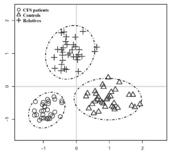
This suggests that there is a pre-disposition via the salivary microbiome for ME/CFS. The salivary microbiome is formed by kisses, and just being in the same living space (this was demonstrated in a recent study where the microbiome of family pets were transferred to the children [2021] [2021])

It is nice to see this clearly demonstrated. This has been reported prior Chronic fatigue syndrome patients have alterations in their oral microbiome composition and function. [Sep 2018]

In prior posts, over the last 7 years, I have flagged this issue:

What are the changes in the mouth bacteria (with links to what modifies these bacteria)

Bottom Line

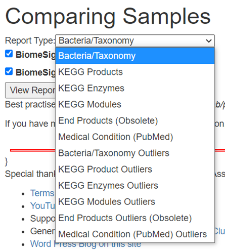
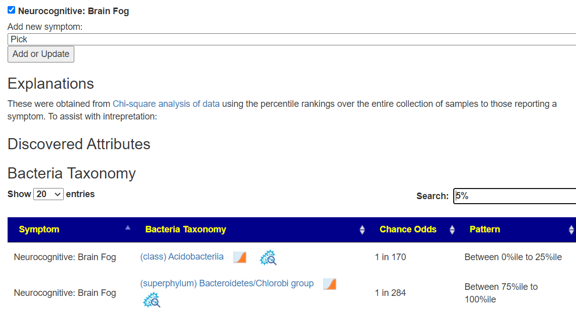
The authors asked “if they are related to any of the several secondary symptoms“, the answer is yes. For several years we have demonstrated that on Microbiome Prescription with citizen science.

https://microbiomeprescription.com/Library/CitizenScience?pvalue=0.01&symptomIds=289

Similarly, “if the microbial composition changes are cause or consequence of the onset of CFS/ME” can be answered as: Some transitory event (i.e. infection, stress, vaccination) causes a change of the microbiome composition. This altered composition takes on a life of its own and cascade into ME/CFS.

My own experience, and some others, are that by altering the microbiome composition back towards normal, ME/CFS improves or disappears.

The unfortunate aspect is that it cannot be handled in ‘one drug solves it’ or a pro-forma cookbook by medical professionals. Every individual has a different microbiome shift with items suggested for one ME/CFS person being contraindicated for a different ME/CFS person based on their 16s determined microbiome and using the artificial intelligence used for suggestions on Microbiome Prescription.

My expectations are that they will be no traction towards treating ME/CFS by conventional researchers. The reason is simple — the ground rules for studies and processes will send them into cascading challenges which required “stepping outside” of establish processes to get traction. This has been described as the “Dogma of Conformity”.

A review of a new CFS/ME Hypothesis

A reader asked for my thoughts on “Pathophysiology of skeletal muscle disturbances in Myalgic Encephalomyelitis/Chronic Fatigue Syndrome (ME/CFS)” 21 Apr 2021. With over 20 years of reading many many hypothesis of ME/CFS, I have learnt to look for earlier indicators that it will be unlikely.

First, “skeletal muscle disturbances” applies to a subset of ME/CFS only. The second item is simple – is this a new hypothesis or an old one-recycled? What I found was:

What is the new hypothesis?

The author’s earlier title was “A Unifying Hypothesis of the Pathophysiology of Myalgic Encephalomyelitis/Chronic Fatigue Syndrome (ME/CFS): Recognitions from the finding of autoantibodies against ß2-adrenergic receptors” [2020] There is no reference to acylcarnitine in the document – this is a red flag for an under-researched paper. You want to cite and discuss prior papers on the same topic.

“sympathetic overactivity in the presence of vascular dysregulation by ß2AdR dysfunction causes predominance of vasoconstrictor influences in brain and skeletal muscles,”

The conclusion is below and amounts (IMHO) to a bunch of hand-waving. The key factor is that the hypothesis is really not testable (hence authors get credit for a publication, suffers of ME/CFS gets nothing).

Considering all the potential predisposing factors we think that in the presence of a high sympathetic tone dysfunction of the ß2AdR could play an essential role (conditio sine qua non) in the etiology of ME/CFS although secondary and rather complex mechanisms have to get involved and to interact, particularly in the chronification of the disease (Figs. 4 and 5). Dysfunction of the ß2AdR by autoantibodies, mutations and desensitization by chronic stress would favor vascular dysfunction and cause insufficient stimulation of the Na+/K+-ATPase at least as a risk factor together with other precipitating factors. There may be numbers of different, still unknown predispositions.

According to our hypothesis dysfunctions of the ß2AdR or post-receptor mechanisms, of the NHE1, the Na+/K+ATPase, the NCX, the RAAS and the KKS could be causally involved in ME/CFS and should therefore be further investigated. This concept offers potential novel strategies for the treatment of this debilitating disease.

The only use of the word “treatment” is in the last sentence.

Going to the earlier study by the same author, I read “Metabolic changes in ME/CFS are also in line with a concept of hypoxia and ischemia.” — that echoes the old hypercoagulation hypothesis from Dave Berg [1999, 2000]. The key difference is that Berg’s hypothesis was testable (via the ISAC Panel) and a variety of treatments (depending on the results). For myself, it worked and I went into full remission. This was confirmed in a trial but then a series of poorly done studies faded away. I recall one study that tested using baby-aspirin on ME/CFS patients and reported no results, thus dismissing the coagulation hypothesis.

A major factor in Berg’s hypothesis fading was the complexity of multiple treatment plans was more than most ME/CFS physicians wanted to deal with (give me one magic bullet please!) , patients referred to hematologists were tossed back because low grade chronic coagulation was not of interest to them.

How well studied is ß2-adrenergic receptors?

I checked PubMed and found there is only 16 results. Several deal with asthma, and thus I would expect an association to asthma to be in the ME/CFS literature – which I was unable to find

Bottom Line

I would drop this concept into the circular filing cabinet. It lacks any direct test results on ME/CFS patients to support it, offers no treatments. It is full of speculation and “reasoning”, i.e. “Therefore, QTc shortening can be explained only by ß2AdR dysfunction in the presence of intact ß1-adrenergic receptors and a high sympathetic tone.” The use of the phrase “can be explained only” instead of ” could be explained” signals spin-doctoring and salesmanship to me, not careful science.

Piracetam for ME/CFS, Long Haul COVID brain fog

This morning I dropped a note to my physician about immune system activation symptoms from getting my 2nd covid shot. One item cited was suspected activation of coagulation resulting in brain fog. I mention that it disappears for half of the day with 300 mg of Piracetam. I expect an email back asking what is Piracetam?

From Wikipedia, you can get general background. It is NOT to be confused with paracetamol (acetaminophen). It’s sits in a murky space in the US, so while legal, without prescription, it can be hard to find.

It improves brain function for many conditions

  • [2020] After treatment, the quality of life scores was significantly higher than the control group, and the difference was statistically significant (P<0.05). For patients with vascular dementia after cerebral infarction, piracetam combined with nimodipine can improve the cognitive function, improve the quality of life, and have a significant clinical effect.
  • [2021] Although Piracetam is officially recognized as a nootropic, its enhancing effects in the healthy individual’s brain are moderate [104,54]. The racetam molecules are being used across a range of brain disorders, including Alzheimer’s disease, narcolepsy, ADHD, Parkinson’s disease and brain aging [105,106].
  • [2020] Piracetam shifted the balance of mitochondrial fission or fusion toward fusion, which is more favorable for ATP production, which would indirectly benefit synaptic function because of the energy requirements of synapses. 43 A review indicated favorable effects on neurotransmission and neuroplasticity. 44 Another report showed that the increased neurite length after piracetam treatment was accompanied by increased expression of the synaptic marker GAP43, thus improving neural plasticity. 45

In days long ago when I often fly between Seattle and India for work, I used a heparin-piracetam cocktail to reduce risk of DVT. As a side effect, I never experienced jet-lag.

  • [1998] ” The heparin-piracetam complex had a more pronounced anticoagulant and fibrinolytic activities than the individual components.”
  • [1993] “The particular efficacy of 8 g piracetam daily in 3 divided doses at 8-hourly intervals can be attributed to its unique dual mode of action; inhibition of platelet function by inhibition of thromboxane A2 synthetase or antagonism of thromboxane A2 and increased formation of prostaglandin I2, together with a rheological effect involving reduction in blood and plasma viscosity through an increase in cell membrane deformability and a reduction of 30-40% in the plasma concentrations of fibrinogen and von Willebrand’s factor”

This last study is why it is my go to when brain fog occurs. I have a mutation that results in higher levels of fibrinogen.

Coagulation in various forms in ME/CFS was demonstrated in the late 1990’s by David Berg. [Other Berg Links: [A,B, *] .

COVID Coagulation

Doing a search on PubMed for “covid coagulopathy“, we find over 1100 citations. For long haul covid to include persistent coagulation is very probable. It may be due to microbiome alterations or other mechanisms.

Bottom Line

Trying piracetam for brain fog is something to discuss with your MD. You may need to educate him about the contents of this article. The [1993] study was on humans and thus showed safety and also dosage (8 grams/day in 3 dosages).

Piracetam for ME/CFS, Long Haul COVID brain fog

Looking beyond bacteria

An Exploration of the microbiome economy using Microbiome Prescription and a 16s report.

Small intestinal bacterial overgrowth (SIBO)

This is detected by end products produced: Hydrogen and Methane and not specific bacteria.

We can explore for associations of different products of bacteria to symptoms

The challenge – Looking at almost 10,000 items for patterns. This is automated, so let us jump to results.

Predicted Symptoms

Support for multiple samples

Summary

The purpose is to discover associations. Association does not prove causality; but association often reflect severity of symptoms. This gives leads to explore…. Not canned answers…